Every
finding,
measured
against
every earlier
scan.

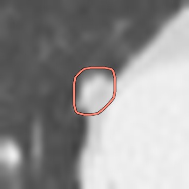
Right upper lobe nodule, 2 years earlier, outlined
2 years earlier
10.2 × 6.8 mmmean 8.5 mm, 388 mm³
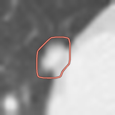
Right upper lobe nodule, 1 year earlier, outlined
1 year earlier+80%
12.0 × 11.0 mmmean 11.5 mm, 697 mm³
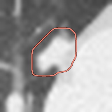
Right upper lobe nodule, Current scan, outlined
Current scan+94%
17.8 × 11.9 mmmean 14.9 mm, 1352 mm³
Slice 65 of 178, the finding's centreAll 3 visits move together. Each image is 42 mm wide.
The sentence Radtechia drafted for this nodule

Right upper lobe nodule: 17.8 × 11.9 mm (mean 14.9 mm), previously 12.0 × 11.0 mm (1 year earlier) and 10.2 × 6.8 mm (2 years earlier). Volume 1352 mm³, +94% in 1 year; volume doubling time 382 days.

Radtechia is an AI assistant for radiologists. It reads a chest CT against every earlier scan, measures what changed and drafts the report. The radiologist checks, edits and signs.

One patient, three scans, nine nodules. Twenty-seven places to look.

Comparing a new CT with every earlier one, finding by finding, by eye, is the heaviest part of the read. Radtechia lays the whole comparison out before the case is opened.
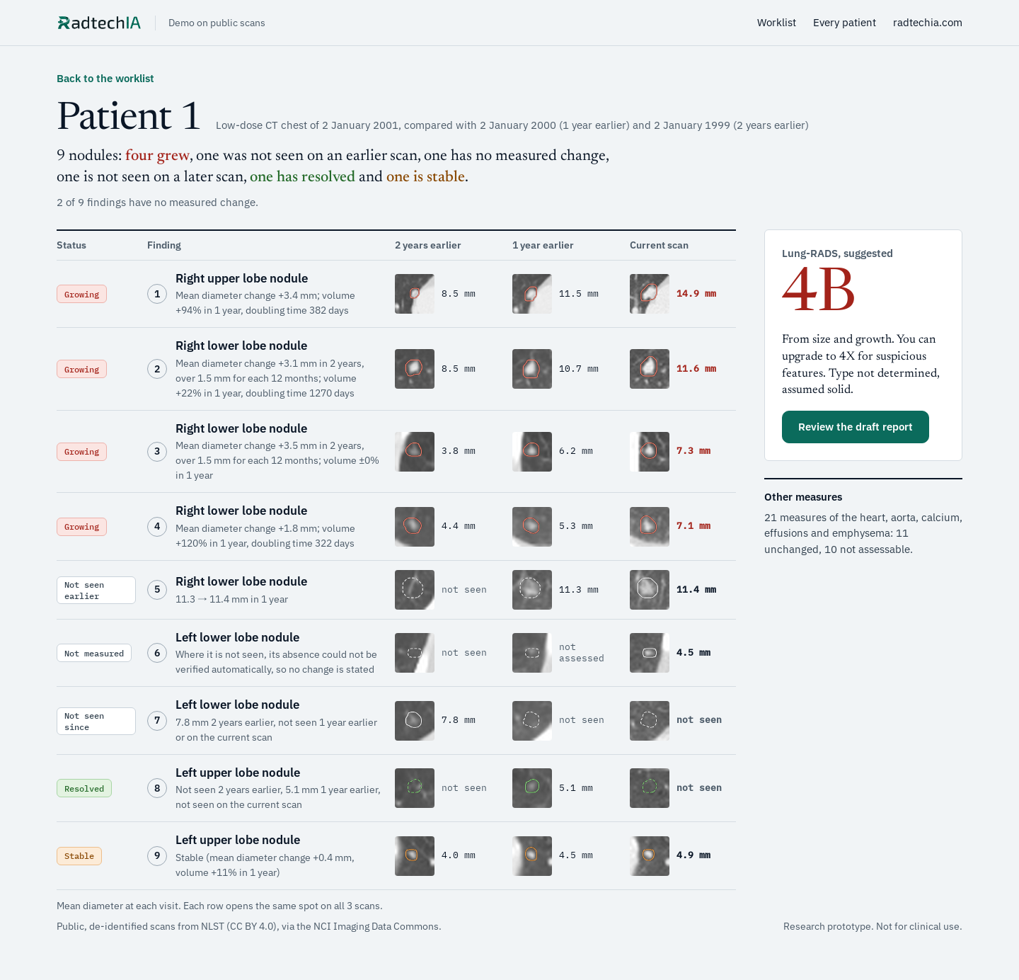
Patient 1: nine nodules, each with its status, its outline and its size on the scan 2 years earlier, 1 year earlier and the current scan. Four are growing, one is stable, one has resolved.
  1. 1Right upper lobe noduleGrowing

    Mean diameter change +3.4 mm; volume +94% in 1 year, doubling time 382 days

    2 years earlier
    8.5 mm
    1 year earlier
    11.5 mm
    Current scan
    14.9 mm
  2. 2Right lower lobe noduleGrowing

    Mean diameter change +3.1 mm in 2 years, over 1.5 mm for each 12 months; volume +22% in 1 year, doubling time 1270 days

    2 years earlier
    8.5 mm
    1 year earlier
    10.7 mm
    Current scan
    11.6 mm
  3. 3Right lower lobe noduleGrowing

    Mean diameter change +3.5 mm in 2 years, over 1.5 mm for each 12 months; volume ±0% in 1 year

    2 years earlier
    3.8 mm
    1 year earlier
    6.2 mm
    Current scan
    7.3 mm
  4. 4Right lower lobe noduleGrowing

    Mean diameter change +1.8 mm; volume +120% in 1 year, doubling time 322 days

    2 years earlier
    4.4 mm
    1 year earlier
    5.3 mm
    Current scan
    7.1 mm
  5. 5Right lower lobe noduleNot seen earlier

    11.3 → 11.4 mm in 1 year

    2 years earlier
    not seen
    1 year earlier
    11.3 mm
    Current scan
    11.4 mm
  6. 6Left lower lobe noduleNot measured

    Where it is not seen, its absence could not be verified automatically, so no change is stated

    2 years earlier
    not seen
    1 year earlier
    not assessed
    Current scan
    4.5 mm
  7. 7Left lower lobe noduleNot seen since

    7.8 mm 2 years earlier, not seen 1 year earlier or on the current scan

    2 years earlier
    7.8 mm
    1 year earlier
    not seen
    Current scan
    not seen
  8. 8Left upper lobe noduleResolved

    Not seen 2 years earlier, 5.1 mm 1 year earlier, not seen on the current scan

    2 years earlier
    not seen
    1 year earlier
    5.1 mm
    Current scan
    not seen
  9. 9Left upper lobe noduleStable

    Stable (mean diameter change +0.4 mm, volume +11% in 1 year)

    2 years earlier
    4.0 mm
    1 year earlier
    4.5 mm
    Current scan
    4.9 mm

2 of 9 findings have no measured change.

Solid outline: measured
Dashed: nothing measured at that visit
Dotted: approximate size

Every number comes from the images.

1. Matched

2 years earlierNot seen
1 year earlier5.1 mm
Current scanNot seen

Each finding is found again on every earlier scan, not only the last one. When nothing is there, the report says so: this one has resolved.

2. Measured

2 years earlier388 mm³
1 year earlier697 mm³
Current scan1352 mm³

Sizes and volumes are taken from the finding's outline on the scan, at every visit, so the change is a measurement and not an impression.

3. Categorised

Growing by Lung-RADS (mean diameter change +3.4 mm).
Lung-RADS 4B, suggested

The change is put into its guideline category, with the rule that produced it beside it. It stays a suggestion until the radiologist approves it.

4. Checked

Where it is not seen, its absence could not be verified automatically, so no change is stated.

Every sentence opens the images it came from. A measurement that fails its own check is never passed off as exact: the draft says what it could not verify.

A draft that reads the way radiologists already write.

Findings and impression, in the usual order. The radiologist reads the draft beside the images, edits what needs editing and signs.

Low-dose CT chest, compared with 2 earlier scans
Draft, ready for your review
Findings

Right upper lobe nodule: 17.8 × 11.9 mm (mean 14.9 mm), previously 12.0 × 11.0 mm (1 year earlier) and 10.2 × 6.8 mm (2 years earlier). Volume 1352 mm³, +94% in 1 year; volume doubling time 382 days. Growing by Lung-RADS (mean diameter change +3.4 mm).

Left upper lobe nodule: 5.1 mm 1 year earlier; resolved.

Impression
  1. 1. Interval growth of the 14.9 mm right upper lobe nodule by mean diameter, volume doubling time 382 days (part of its edge lies against tissue of its own density: volume and doubling time uncertain).
  2. 8. Resolution of the previously seen 5.1 mm left upper lobe nodule.
  3. 10. Lung-RADS 4B (suggested): Diagnostic chest CT with or without contrast; PET/CT may be considered if there is a ≥ 8 mm solid nodule or solid component; tissue sampling; and/or referral for further clinical evaluation. Management depends on clinical evaluation, patient preference, and the probability of malignancy. Type not determined, assumed solid.
Excerpt from the draft for Patient 1. The full draft has nine findings and eleven impression lines.

Lung nodules now. The whole chest next.

Now

Follows lung nodules across every earlier scan, measures their change and drafts the report.

Next

Every finding on a routine chest CT, with or without contrast: lymph nodes, pleura, heart and vessels, upper abdomen.

Measured against radiologists

Each part is scored against radiologists' own readings, on scans kept out of its training. This validation is under way.

Built on public scans

Developed on public, de-identified CT scans, including the National Lung Screening Trial.

An AI radiologist that reads everything.

That is where we are going. We start with one exam, chest CT, and widen the reading one step at a time, as each step is proven.

Nabil Mouadden
CEO and technical lead. PhD candidate in AI, CentraleSupélec.
Kine Nilsen
Sales and partnerships, Norway.

See it read a real case.

Book a demo and we take you through the whole read on public scans: the worklist, what changed, the images at every visit and the draft report.